
This is an excellent clinical question, and the type of question that a clinician encounters almost daily. How should we determine the answer to the question? Rather than just tell you, allow me to go through the thought process that should be applied to this and countless other questions.
First, we must make sure we understand the problem and what factors are important to consider. We must also decide what the important outcomes are and make sure that we are not making crucial decisions based on surrogate outcomes alone. Most mistakes in answering a clinical question like this one actually occur in this step. If we are asking the wrong question, we’ll probably get the wrong answer.
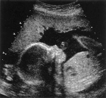
So what is oligohydramnios?
Oligohydramnios has numerous definitions, and this lack of clarity will contribute to confusion in answering our question. It has been defined in many different ways:
- an amniotic fluid index (AFI) less than 5 cm, where AFI is the sum of four vertical pockets from four quadrants
- an amniotic fluid volume (AFV) less than 2 cm in a single deepest pocket (SDP)
- some will define oligohydramnios as less than 5th or 10th%ile of fluid expected for the gestational age
- some trials will consider it appropriate to intervene when the AFI is less than 6 cm, or 8 cm (this is an example of diagnostic drift, but it can significantly impact study results)
- some use purely subjective assessments (it looks normal or it looks abnormal)
- in studies able to measure the fluid objectively, a total volume of less than 200 ml or 500 ml has been suggested.
So first we must decide which of these definitions to use. The truth is, we don’t know how much fluid is “normal.” We can define normal by looking at a cross section of patients and describing the distribution of volumes of fluid in large population of patients, and this has been done. We can then say that anything above or below a certain percentile (the patients on the tails of the bell-shaped curve) are “abnormal,” and this thinking leads to percentile diagnoses, such as oligohydramnios being defined as less than 5th percentile. But that doesn’t necessarily mean that having that amount of fluid is associated with negative outcomes. So abnormal, in that context, may not necessarily be “bad.”
Part of the problem with defining oligohydramnios by either the AFI < 5 cm or the AFV < 2 cm is that the techniques to measure fluid with ultrasound just aren’t that good. The largest study to compare ultrasound methods to objective measurements of amniotic fluid (like dye dilution studies or direct measurements) found that the AFI method only had a sensitivity of 10% and a specificity of 96% while the AFV method (single deepest pocket) only had a sensitivity of 5% and a specificity of 98%. This makes both tests very unreliable for predicting how much fluid is present. Other studies have shown that these predictive values don’t improve even when a variety of techniques are combined.
So our techniques for measuring amniotic fluid just aren’t that good. They have low predictive value and significant unreliability. They also have poor reproducibility. One lesson from this is that sufficient pretest probability should be present before any type of ultrasound-based measurement of amniotic fluid is made, else the result is next to meaningless.
A better way to define how much fluid is abnormal is by correlating the level of measured fluid with negative pregnancy outcomes, rather than as a subset of a normal distribution of amniotic fluid volumes across a population. In other words, our ability to predict how much fluid is actually in the uterus is very poor, but do the poor measurements we take correlate with good or bad outcomes in a clinically useful way? Before we look for answers to that question, we will stumble across one more related issue. Ultrasonographers are trained to exclude pockets of fluid that contain umbilical cord from their measurements; in the past, this was done only with gray scale ultrasound, but most current ultrasonographers can do a better job of excluding cord by using color doppler. Studies have shown that the rate of over-diagnosis of oligohydramnios using color doppler is about 20% compared to using gray scale alone. This is important because original studies that tried to determine which levels of amniotic fluid were associated with abnormal pregnancy outcomes did not use the color doppler method.
There are only a few studies that have looked at dye-determined, objectively measured oligohydramnios which also evaluated fetal outcomes. One found no difference in meconium-stained fluid, variable decelerations, or low Apgars; another found no difference in fetal pH; and another found no difference in a variety of intrapartum outcomes studied, including late and variable decelelerations, IUGR, mode of delivery, NICU admission, etc. These facts are important because it would seem that there is no scientific evidence for most of the intrapartum negatives that are anecdotally associated with low amniotic fluid.
The literature related to an ultrasound-derived AFI <5 and adverse outcomes is mixed to say the least. The classic paper by Rutherford et al. from 1987 that largely established the current fears of oligohydramnios is itself plagued with methodological problems, but it armed perinatologists with a new tool in the form of AFI measurement and a reason to be worried about it. This retrospective chart review of 353 women found just 27 with an AFI of less than 5 cm. The main reason these ultrasounds were done were because of “postdate” pregnancies, many of whom did not have “good dates.” Not surprisingly, they found that the women with low fluid were more likely to have meconium-stained fluid, intrapartum distress, etc. But is this because they were “postdate” or because they had a low amount of amniotic fluid? In other words, do the fetal distress and low fluid have a common cause (like chronic uteroplacental insufficiency), or is the low fluid itself causing the distress? The main problem with this influential paper is that its findings are not generalizable. The finding of low fluid in these pregnancies may have just been a way of better identifying more severe postdatism; but nothing in the study implies that the results are applicable to a well-dated pregnancy, say at 30-weeks-gestation.
Chauhan et al. conducted a meta-analysis of 18 trials that evaluated the relationship between low fluid and adverse outcomes. They concluded that an AFI of less than 5 was associated with an increased risk of cesarean delivery for fetal distress and an increased risk of 5 minutes Apgar scores less than 7, but no evidence of true distress, such as fetal acidosis. This finding is interesting and raises the question of whether bias related to the providers’ knowledge of the presence of oligohydramnios might have affected their management of the labors. How many women were perhaps unnecessarily induced and unnecessarily delivered by cesarean because the provider was anticipating a negative outcome in a “high-risk” pregnancy, even though studies that look at objective measurements of oligohydramnios fail to see such associations? How well can the studies in this meta-analysis be trusted since ultrasound is so poor at detecting oligohydramnios in the first place? Essentially, studies have shown that if you have oligohydramnios, you are more likely to be delivered by cesarean for “fetal distress” by your obstetrician, but not more likely to have actual, objective fetal distress.
Another review of studies conducted in 2016 by Rabie et al. identified 15 trials to be analyzed. They concluded that among low-risk pregnancies, women with isolated oligohydramnios were more likely to have neonates with meconium aspiration syndrome, to have a cesarean for fetal distress, and to have a NICU admission. Patients in the high risk group were more likely to have infants with low birth weight, but all other metrics were the same, including Apgar scores, NICU admissions, meconium-stained fluid, and cesarean delivery. So once again, the issue is likely that the oligohydramnios is a result of the same underlying pathology (and the presence of the diagnosis itself may bias providers). In other words, why was amniotic fluid being checked in the low-risk group of women? Largely because of postdatism; we expect higher rates of meconium aspiration syndrome, NICU admissions, and cesareans in that group. Why was the amniotic fluid being checked in the high-risk women? For all sorts of reasons, including things that affect placental blood flow like hypertension and diabetes, and given the underlying pathology behind those types of maternal conditions, when uteroplacental insufficiency occurs, we often see oligohydramnios and growth restriction. But the addition of oligohydramnios to all of the other problems of the pregnancy did not make the pregnancy (or the neonate) any worse off, though it was associated with the presence of growth restriction.
Chauhan et al., in other study, showed that if the obstetrician knew that a patient had oligohydramnios at the time of admission for labor, the patient was more likely to undergo a cesarean delivery for fetal distress; but the fetal outcome was not improved by this knowledge or action. This example of the framing effect clouds all of the non-blinded studies done on this issue, particularly in regards to the outcome of “cesarean for fetal distress,” and it also shows one of the very real iatrogenic harms of over-testing with ultrasound.
The summary of this data, particularly when the individual trials are looked at, is that there is no data that supports the idea that objective outcomes are different in pregnancies that have oligohydramnios determined by ultrasound versus those that do not.
How should we measure oligohydramnios?
When you understand how inconsequential oligohydramnios is, and that the risks of over-diagnosis outweigh the benefits, then the answer intuitively is that we should use the method of ultrasound determination that labels the fewest women as having oligohydramnios, which happens to be the AFV method (single deepest pocket < 2 cm). This has been studied in several modern trials.
- Alfirevic et al. conducted a randomized controlled trial of 500 women comparing AFI to AFV in 1997. They found that a woman was 4.5 times as likely to be diagnosed with oligohydramnios if AFI was used rather than AFV, and this meant more inductions and more monitoring, with a trend toward more cesareans (18.8% vs 13.2%). Other outcomes were similar.
- Magann et al. also did an RCT in 2004 comparing AFI vs AFV as a component of a biophysical profile. They similarly found that 2.84 times as many women were labeled as having oligohydramnios when AFI was used, but neonatal outcomes were the same.
- Moses et al., also in 2004, performed a RCT comparing the two methods. They found that about 3 times as many women were labeled as having low fluid with the AFI method, but no other differences in outcomes. Interestingly, they also found that neither method was able to accurately identify the women who would subsequently undergo an amnioinfusion for fetal distress, who would have variable or late decelerations, fetal distress, cesarean for fetal distress, or NICU admission (that is, both tests were worthless).
- Finally, also in 2004, Chauhan et al. randomized 1080 women to the two methods. Again, they found that more women were labeled as having oligohydramnios with the AFI method (17% vs 10%). There otherwise were no differences in outcomes except a trend towards more cesareans in the group over-diagnosed with oligohydramnios.
So if you’re still planning on measuring amniotic fluid at this point, the take-away is that you should be using the single deepest pocket method, in order to prevent iatrogenic harm. Unfortunately, the rates of utilization of AFI (instead of SDP) remain very high. One wonders if this is due to the economic incentives of over-diagnosis.
Do we care about oligohydramnios then?
If oligohydramnios is seemingly unimportant, then why even look for it? And if abnormal, why try to treat it (with hydration for example)? I have previously discussed the PORTO study in relation to IUGR, but the study is useful for thinking about oligohydramnios as well. The PORTO study found that oligohydramnios was a predictor of an adverse perinatal outcome only when associated with an estimated fetal weight (EFW) less than the 3rd percentile. This observation should provide some context for what the clinical utility of oligohydramnios truly is (apart from its use in the BPP). If oligohydramnios is important, it is important because it is a sign of either ruptured membranes, significant uteroplacental insufficiency (UPI), or some relatively rare condition like Potter’s sequence.
If we have ruled out ruptured membranes, then we must assess for evidence of UPI. If there is chronic UPI, then a fetus will begin to shunt blood away from the splanchnic circulation and the renal arteries to favor the brain and heart. When this occurs, an immediate consequence may be decreased urinary production. Since the whole amniotic fluid volume is turned over daily, then less fetal urine can be a first sign of decreased fetal perfusion. Eventually, this selective shunting of blood will also result in a small abdominal circumference (as the liver becomes relatively smaller) and, finally, asymmetric growth restriction. By the time this IUGR becomes severe (<3%ile), then the rate of adverse outcomes increases. Along the way, or perhaps as a late finding, we might also observe changes in the umbilical artery velocimetry, as relative placental resistance increases. These changes in the umbilical artery doppler profile are also associated with an increased risk of adverse outcomes.
So that’s it. We care about oligohydramnios only because it is an indirect sign than there might be UPI, which is ultimately the cause of the adverse outcomes (not cord compression, placental compression, or other nonsense). So what if the fluid is low but the fetus is growing well? In this case, it may be a transient issue that resolves, an aberrant reading, a normal variation, or a sign of things to come (that is, the first sign of a placenta at risk). The only way to know this is to recheck the fluid and see if there is a persistence of the abnormality, and if that persistence eventually becomes associated with growth restriction.
What about the BPP? The biophysical profile is a useful tool for determining fetal well-being. Yet, as noted above, studies don’t seem to show a difference in BPP predictive value dependent upon which method of measuring amniotic fluid is used. This means that a lot fewer abnormal BPPs will happen if SDP is utilized rather than AFI, but with the same predictive value for abnormal outcomes. Usually, when amniotic fluid alone makes the difference in a BPP, it is related to postdatism, and in this regard, measuring amniotic fluid as part of the BPP should likely remain a part of the BPP test. Currently, BPPs are over-utilized in clinical practice (due to diagnostic drift) and, if the test is conducted with AFI rather than SDP, then a significant number of over-diagnoses are likely occurring.
Does it matter then if we correct the amniotic fluid volume by maternal hydration?
You have probably already guessed the answer to this question: No. There is simply no scientific evidence that shows that iatrogenically correcting amniotic fluid volume is valuable. Let’s say that the low fluid is due to ruptured membranes; will increasing it be helpful? No. What about if it is transient or has some cause related to something other than chronic UPI? Will it be helpful then to make the number on the ultrasound report greater? No. Well, what if it is due to chronic UPI? Won’t it be helpful to increase the amniotic fluid and therefore increase placental perfusion with forced hydration? I think that’s the spirit of the idea, but there is simply no scientific evidence that increasing maternal hydration for a short time (a few hours or perhaps days) has any impact on chronic UPI. The uteroplacental interface is not a pump that needs to be primed and then growth restriction and fetal oxygenation will be improved thereafter. So even if maternal hydration were to improve the ultrasound diagnosis of oligohydramnios, it might actually have a deleterious effect on the gestation since it might give false reassurance that there is an appropriately working placenta, rather than preserve the early warning system of severe growth restriction and increasing placental resistance.
The point is, even if maternal hydration improved the diagnosis of oligohydramnios, we need to know if it will also improve fetal outcomes. This is an important point. The ultrasound measurement of amniotic fluid is a surrogate marker, and a very, very poor one. Remember how badly ultrasound is at actually measuring the true amount of amniotic fluid in the first place. Often, poor measurements come just from pressing too hard on the maternal abdomen or an incorrect angle of the probe. It’s a very poor test to base important decisions on, particularly in cases of isolated oligohydramnios. When oligohydramnios is the only abnormal finding, then the pretest probability is very poor, and therefore, even in the presence of a positive finding, the positive predictive value is also exceedingly poor. The lesson again here is that the test shouldn’t be done when there isn’t sufficient pretest probability to do so (like a specific maternal medical condition associated with UPI), and when it is done, it should be performed using the SDP method.
So does maternal hydration improve oligohydramnios?
Well, I don’t care. It’s not relevant. But if you must know, you can read this low-quality meta-analysis about the subject that concludes that oral hydration is better than intravenous hydration and that hypotonic solutions are better than isotonic solutions. I’ll say now that I took the long way of providing that answer because the more direct way (searching PubMed for the latest meta-analysis) would have given you the wrong idea. Most folks likely would have only read the abstract, and most would not have answered these previous fundamental questions.
But by now, hopefully you realize the house of cards that this meta-analysis represents. Of the 16 papers reviewed, none of the papers used the SDP method, including the ten published after 2004 (the year it became obvious that AFI should be abandoned). Only one looked at outcomes other than the ultrasound measurement of amniotic fluid volumes, and that paper, of course, found a higher rate of cesarean in the oligohydramnios group. Yet this study, Bangladeshian paper about oral hydration, should have been excluded due to its low quality, low number, and 11% fetal death rate.
Ten of the 16 papers had 25 or fewer patients enrolled, making the majority of included studies statistically worthless. The highest quality and largest study in the mix is this paper from 2014 that looked at whether IV administration of 2 L of hypotonic fluid prior to attempted external cephalic version impacted the success rate of the procedure. It did not. None of the papers accounted for the margin in error in ultrasound measurement of the fluid and most of the authors didn’t consider blinding, randomization, or placebo-control as important things to do in a study.
In short, 16 low quality studies don’t make one good one. Some of the small studies showing success of maternal hydration contain conclusions like this one: “Since it caused no complications for the mother and the fetus, it can be used as an effective method in management of oligohydramnios.” And isn’t this the problem with “scientific” publications? This Iranian study of 20 patients, using inappropriate statistical methods, unpowered to detect serious complications (like maternal heart failure or water intoxication), with no evidence of improved neonatal outcomes, no surrogate markers measured more than 90 minutes after the intervention (what was the fluid the next day or next week?), using the wrong method for detecting fluid volume (the AFI), producing a clinically insignificant effect (1.5 cm increase of AFI), and published in the prestigious Journal of the Caring Sciences (an open access journal), is considered science. What’s more, they have the hubris to claim that this is “managing” oligohydramnios.
Yes, this paper was one of the 16 trials included in the previously cited, open-access published meta-analysis, where the lead author cited himself 17 times in the bibliography, with such irrelevant publications as Fertility rate and subsequent pregnancy outcomes after conservative surgical techniques in postpartum hemorrhage: 15 years of literature. Are you seeing the real problem? These studies and publications shouldn’t have been published in the first place, but the publishers are making money and the “researchers” are going to get published and are going to increase how many times their publications have been cited even if they have to cite them themselves and pay for it to be printed. Somewhere, some “academician” advanced their careers (and harmed patients) with such garbage.
But I’m on a rant. I’m sure the editor of PLOS One, which publishes 70% of all received manuscripts at the tune of 85 papers each and every day, saw some value in the paper other than the $1,495 publication fee (yes, that’s $127,075 per day). No wonder some people joke that the ‘L’ should be dropped from the journal’s title.
Well, anyway, the take-home points are these:
- Oligohydramnios is only important when used as a warning sign for ruptured membranes, chronic uteroplacental insufficiency, or other rare conditions like urinary outlet obstruction or Potter’s sequence.
- We should measure amniotic fluid only when necessary and only using the single deepest pocket method, where normal is when any pocket is greater than 2 cm.
- Don’t recommend hydration (and certainly don’t hospitalize someone for IV hydration) just because she has oligohydramnios.
- Stop thinking that isolated oligohydramnios is an ominous sign, and stop doing unnecessary cesareans.
- Stop reading nonsense published in PLOS One and other for profit paper-mills.